International Journal of Veterinary Science and Research
Cervico vaginal prolapse correction and management by using bilateral configuration of sterile synthetic plastic materials in local breed cow
Jiregna Dugassa Kitessa1* and Kisi Bekele Terefe2
2DAH, BVSc, Veterinary Clinical Practitioner at Hiddi Veterinary Clinic, Ada’a District, East Shoa Zone, Oromia, Ethiopia
Cite this as
Kitessa JD, Terefe KB (2021) Cervico vaginal prolapse correction and management by using bilateral configuration of sterile synthetic plastic materials in local breed cow. Int J Vet Sci Res 7(2): 073-076. DOI: 10.17352/ijvsr.000083Background: Cervico vaginal prolapsed is one of the most common disorders of the reproductive organs at the end of pregnancy and rarely after.
Objective: Description of the surgical management of cervico vaginal prolapsed in normal delivery.
Animals: Emaciated, local breed cow that was kept under an extensive management system.
Study design: Case report
Study methods: Patient history and clinical findings, diagnosis, treatment
Outcomes: After frequent follow up and well provision of post operative care, the patient was recovered.
Conclusion: Cervical-vaginal prolapsed can lead to infertility and sterility unless it is treated early, before it worsens. The use of sterile rectangular plastic material to prevent recurrence on the bilateral sides of the vulvar lips together with adequate sutures appears promising and is highly recommended for similar cases.
Introduction
Uterine prolapse is a fairly common condition on dairy farms and it may involve the complete prolapse of the uterus, vagina and cervix [1]. Cervix and Vaginal Prolapse (CVP) is one of the common reproductive disorders of ruminants in late gestation and is rare post parturition [2,3]. It is often found in large and small ruminants such as cows, buffaloes and sheep. However, the incidence and symptoms have been extensively elaborated in cows [4]. According to some authors, the incidence of reproductive tract prolapse in cattle ranges between 1-2% [5]. It is also hypothesized as protrusion of all or part of the prolapsed vagina through the vulva is a commonly encountered case in pluriparus cows [6].
Although cervicovaginal prolapsed is a reproductive disease that leads to decrement of production and productivity, the exact cause of cervico-vagianal disorder in cattle have not been well established [7]. However, there are various predisposing factors associated with cervico-vaginal prolapse. Among these; uterine atony, dystocia and hypocalcaemia [8]. It should always be treated as a veterinary emergency in order o have good prognosis [9]. This is because early intervention and repair of enlarged uterine tissues and associated tissues is important in ensuring the survival, early rehabilitation and fertility of the reproductive tract. However, poor or delayed intervention may result in bleeding and contamination resulting in infection, shock, gangrene formation and death [8].
The management and correction of a prolapsed cervico-vagina usually involves, disinfection and washing the prolapsed organ, reduction in size of edematous with glycerol, replacement of organ and application of stay sutures such as Buhner’s sutures [10-12]. The purpose of this surgical case report is to explain the correction and management of cervico vaginal prolapse in a local breed cow following parturition in the field before 12 hours.
Description of the case
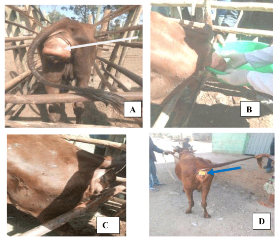
Case history and clinical examination: A six-year-old native cow having parturition prior to twelve-hour was admitted by the owner to the Dire veterinary Clinic, Ada’a District, East Showa zone, Ethiopia. According to the owner, the cow suffered a loss of mass through the vaginal opening immediately after calving. During the expulsion of the calf, the owner assisted carefully and manually. The customer also reported that the pieces that were dropped gradually increased in length and size. The owner tried to help the cow, but the business continued. A careful physical examination revealed prolapse of the vagina and cervix without additional organs such as the bladder (Figure 1A). There is also some dried and dead tissue outside and hanging masses. Based on a thorough history and physical examination, the cow was diagnosed with cervico-vaginal prolapse and decided to undergo surgical correction and management by using the bilateral configuration of sterile synthetic plastic materials in local breed cow.
Preoperative preparations, anesthesia and animal control
The cow was restrained in a standing position as shown in (Figure 1A) and caudal epidural anaesthesia was performed using 2% Lidocaine HCl (2% lidocaine hydrochloride, jeil pharma. co. Ltd., Korea) @0.22mg/kg in the intercoccygeal space. The prolapsed mass was lifted upward above the level of ischial arch and then mass was debrided, cleaned and washed with warm, sterile water followed by the sterile physiological saline solution to remove the debris (Figure 1B) and poured with cold water to decrease a few edemas and size of the mass before relocation.
Correction and management
After the preformed mass was evenly prepared, they were raised with both hands and inserted into the pelvic cavity through the opening of the vagina with the thumb fist starting from the base (Figure 1C). Standing suture (Figure 1D) was made the vulvar lips apart from the vaginal orifice by bilateral configuring of sterilized slightly rectangular synthetic plastic material after boiling and cutting to the appropriate size as shown in figure D. The cut rectangular strip was disinfected withdiluted chlorhexine solution before application. Then both synthetic plastic strips was configured bilaterally and fixed in place with horizontal mattress suture by using silk 2-0 size to prevent recurrence. Finally, after 6 days of complete regression, the suture material was removed.
Post operative care of dam and outcome
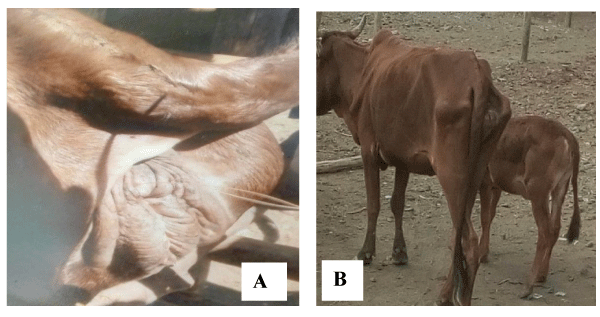
The cow was given penicillin (24mg/kg) and dihydrostreptomycin sulphate (30mg/kg) (Pen &Strep® Norbrook UK) intramuscularly for 3 consecutive days. The owner was also recommended and advised to provide well feed and management in addition to attending and inspecting the recurrence as well as any discomfort felt such as during urination. For topical application at vulvar lips with 5% Povidin iodine solution was recommended. The suture material was removed at day six post surgical reproductive management (Figure 2A) and completely recovered.
During continuous monitoring, the body condition of the cow decreased slightly due to the decrease in feed availability, but it was observed that her calf grew in good body condition (Figure 2B).
Discussion
Genital prolapse, including cervical and vaginal prolapse in ruminants, is considered as an urgent maternal disease that requires immediate intervention before further complications ensue [13]. Because the delay of the provision of management and correction leads to the necrosis, infection and the rupture of prolapsed mass. It has been reported that vaginal prolapsed is rare after 48 to 72 hours after birth. The aim is to replace the organ with the method that can hold it in a fixed position [5]. This is in line with the guidelines in the present case as retaining stitches have been placed by using bilateral configuration of sterile srips of plastic material. On the other hand, vascular compromise, trauma and faecal contamination may also increase toxin intake across the cervico-vaginal mucosa. However, careful removal of these materials, after soaking with warm dilute antiseptic solution is usually successful causing only minor capillary bleeding [14].
The present case of cervicovaginal prolapsed can be traced back to the recent calving due to forced pulling [4]. Treatment of an organ prolapsed invariably leads to tenesmus, so that light epidural anesthesia is required [6]. This in turn looks like epidural analgesia has been performed using a local anesthetic to relieve pain and tenesmus.In the present clinical case, the modified Bühner’s technique was not applied due to lack of the tape and it’s needle at the field condition and rather retention suture was employed by using available local material after sterilizing, cutting and configuring bilateral to vulva lips in order to prevent tearing from tension of suture material and thereby avoid recurrence. It was found to be very satisfactory in preventing recurrence of the prolapsed mass and therefore is recommended as an alternative technique, particularly in developing countries where farmers cannot afford costly repeated treatment of their livestock [15]. The advantages of this modified technique over the standard Bühner technique include sufficient space (between the suture knot and the ventral vulvar commissure) for easy urination, no need to make and sew the incisions above and below the vulva, the suture can be loosened and reapplied by the owner himself as needed, without the need for additional labor and other sophisticated instruments, and without anatomical distortions or physiological defects in the vulvar area.
Conclusion and recommendations
Cervico vaginal prolapse is one of the reproductive emergencies of cows due to different factors which needs immediate surgical; treatment as it hampers the production and productivity either periparturion or immediately after few hours of parturition. However the severity depends on the duration and level of contamination besides the size of prolapsed mass. Thus, in this special surgical case report, correction and management were effectively addressed by putting stay sutures combined with bilateral configuration of sterile synthetic plastic strip material to the vulvar lips for a short period of time to prevent recurrence. Finally, the cow was completely relieved and alive. Therefore, the authors recommend that similar can be managed before they escalate immediately, and possibly with this new method especially in countries where resources are scant and veterinary services coverage are not grass root level.
Both authors warmly extend their deep and heartfelt gratitude to veterinarians and technicians of Dirre Veterinary clinic for their materials and provisions of inputs during case handling.
Author contributions
In addition to case management, both authors contributed to the conception and design of the study. The correction of the case and the writing of the manuscript will be in charge of JD, while KB assisted in the processing of the case and commented on the previous versions and the draft of the manuscript. Finally, both authors read and approved the final manuscript.
Availability of data and materials
All data generated or analyzed during this study are included in the manuscript for publication.
Ethics approval and consent to participate
Ethical approval was waived by the local Ethics Committee of University. An in view of the survey report nature of the study and all the procedures being performed were part of the routine care and procedures.
- Divers TJ, Peek S (2007) Rebhun's diseases of dairy cattle. Elsevier Health Sciences 400-404. Link: https://bit.ly/3iHylUf
- Raidurg R (2014) Surgical management of cervico-vaginal prolapse in a hallikar cow. Intas Polivet 15: 470-471. Link: https://bit.ly/37TcaEt
- Noakes ED, Parkinson TJ, England GC (2009) Veterinary Reproduction and Obstetrics. 9th edn. W.B Saunders Company, Philadelphia 146-153. Link: https://bit.ly/3jO2O2h
- Yimer N, Syamira SZ, Rosnina Y, Wahid H, Sarsaifi K, et al.(2016)Recurrent vaginal prolapse in a postpartum river buffalo and its management. Buffalo Bulletin 35: 529-534. Link: https://bit.ly/37Eqssi
- Yotov ST, Atanasov A, Antonov A, Karadaev M (2013) Post oestral vaginal prolapse in a non-pregnant heifer (a case report). Trakia Journal of Sciences 11: 95-101. Link: https://bit.ly/3yL5Ydc
- Tyagi RS, Singh J (2002) A Textbook of Ruminant Surgery: 1st Edn. Blackwell publis.
- Noakes DE, Perkinson TJ, England GW (2001) Post Parturient Prolapse of the Uterus. Arthur’s Veterinary Reproduction and Obstetrics. Saunders 333-338.
- Andrews AH, Blowey RW, Boyd H, Eddy RG (2008) Bovine medicine: diseases and husbandry of cattle. John Wiley and Sons514-515. Link: https://bit.ly/3CLVHQo
- Yadav DS, Choudhary R, Shakkarpude J, Gautam M (2014) Postpartum uterine prolapse and its therapeutic management in a buffalo. Intas Polivet 15: 426-428. Link: https://bit.ly/37BCkuX
- Makhdoomi DM, Gazi MA, Batacharya HK, Sofi KA (2014) Surgical management of uterine prolapse in a cow. Intas Polivet 15: 410-412. Link: https://bit.ly/2VJAczh
- Patel RK, Patel N, Singh N, Singh P (2016) Successful management of rectal prolapse in a mule (Equus mulus). J Livestock Sci7: 35-37. Link: https://bit.ly/3m1cDfU
- Simon MS, Gupta C, Ramprabhu R, Prathaban S (2014) Postpartum uterine prolapse and its management in a cow. Intas Polivet 15: 415-417. Link: https://bit.ly/3jPMFcC
- Murphy AM, Dobson H (2002) “Predisposition, Subsequent Fertility and Mortality of Cows with Uterine Prolapsed. Veterinary Record 151: 733-735. Link: https://bit.ly/2UhG9CK
- Senthil KA, Yasotha A (2015) Correction and Management of Total Uterine Prolapse in A Crossbred Cow. Journ Agric Vet Sci 8: 14-16. Link: https://bit.ly/3sdFbDR
- Prakash S, Selvaraju M, Manokaran S, Ravikumar K, Palanisamy M (2016) Obstetrical management of total uterine prolapse in a Kangeyam heifer. Int J Sci Environ Technol 5: 1952–1954. Link: https://bit.ly/3xJktx0
Article Alerts
Subscribe to our articles alerts and stay tuned.
 This work is licensed under a Creative Commons Attribution 4.0 International License.


 Save to Mendeley
